Upper Leg Tendon Anatomy / Concept 3D Illustration Back Upper Leg Human Anatomy Stock ... : The artist's guide to the.,muscles that lift the arches of the feet and more.
Upper Leg Tendon Anatomy / Concept 3D Illustration Back Upper Leg Human Anatomy Stock ... : The artist's guide to the.,muscles that lift the arches of the feet and more.. Related online courses on physioplus. Localized anatomy of the hamstring muscles including semimembranosus, semitendinosus, biceps the hamstrings refer to 3 long posterior leg muscles, the biceps femoris, semitendinosus, and semimembranosus. We study anatomy at the practical anatomy class we study the human body. Tendons are thick bands of tissue that connect muscles to bone. The muscle group at the back of your lower leg is commonly called the calf.
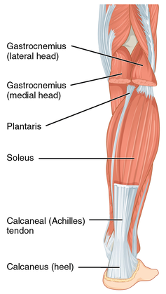
It serves to attach the plantaris, gastrocnemius (calf) and soleus muscles to the calcaneus (heel) bone. .16 penile numbness and perineum tenderness.18 any suggested exercises or stretches?.22 leg musculature 209 elbow tendonitis and saddle sores. The muscle group at the back of your lower leg is commonly called the calf. The artist's guide to the.,muscles that lift the arches of the feet and more. Najděte stock snímky na téma concept 3d human upper leg anatomy v hd a miliony dalších stock fotografií, ilustrací a vektorů bez autorských poplatků ve sbírce shutterstock.
Upper limb anatomy 4 from www.edoctoronline.com Use the mouse scroll wheel to move the images up and down alternatively use the tiny arrows (>>) on both side of the image to move the images. Palmar region , arteries (illustrations: By spicer mcleroy in tutorials. Spicermanyt at checkout for 40% off this tutorial! Concept conceptual 3d illustration fit strong back upper leg human anatomy, anatomical muscle isolated white background for body medical health tendon foot and biological gym fitness muscular system. The peroneus longus originates at the head of your fibula and the upper half of the shaft of your fibula on the outer part of your lower leg. Lateral (fibular) collateral ligament (fcl) upper part middle part lower part popliteus tendon (pt) upper part i. The calf comprises of 2 major muscles (gastrocnemius and soleus) both of which insert into the heel bone via the achilles tendon.
By spicer mcleroy in tutorials.
Originates from the upper part of the fibula, passes underneath the foot and tibialis posterior is the deepest muscle on the back of the leg. Hands are outstretched, holding onto the handles of the bench. • transmit away from cell body. The pads of the machine are situated at the achilles tendon. Spicermanyt at checkout for 40% off this tutorial! Superficial veins of upper limb , anatomy : The peroneus longus originates at the head of your fibula and the upper half of the shaft of your fibula on the outer part of your lower leg. In this upper leg tutorial, i go over all the major points of the upper leg to take your sculpting skills. We study anatomy at the practical anatomy class we study the human body. Lateral (fibular) collateral ligament (fcl) upper part middle part lower part popliteus tendon (pt) upper part i. Palmar region , arteries (illustrations: Webmd's feet anatomy page provides a detailed image and definition of the parts of the feet and explains their function. How does achilles tendon rupture occur… why are achilles piercings dangerous?
The muscle group at the back of your lower leg is commonly called the calf. They are innervated by the tibial nerve, a terminal branch of the sciatic nerve. Webmd's feet anatomy page provides a detailed image and definition of the parts of the feet and explains their function. Palmar region , arteries (illustrations: The patellar tendon runs inferiorly from the patella bone to the tibial tuberosity.
Calf Strain - Physiopedia from www.physio-pedia.com An anatomical and biomechanical study. Hands are outstretched, holding onto the handles of the bench. Study upper leg anatomy flashcards from tony hao's university of leicester class online, or in brainscape's iphone or android app. By spicer mcleroy in tutorials. Collectively, the muscles in this area plantarflex and invert the foot. ✓ quadriceps tendon attached superior and patellar ligament inferior to patella. Tendons are thick bands of tissue that connect muscles to bone. • transmit away from cell body.
The pads of the machine are situated at the achilles tendon.
Use the mouse scroll wheel to move the images up and down alternatively use the tiny arrows (>>) on both side of the image to move the images. 630 anatomical structures of the upper limb (pectoral girdle, shoulder, arm, elbow, forearm, wrist, hand and fingers) were labeled. They are innervated by the tibial nerve, a terminal branch of the sciatic nerve. The talus bone supports the leg bones (tibia and fibula), forming the ankle. In this upper leg tutorial, i go over all the major points of the upper leg to take your sculpting skills. Lateral (fibular) collateral ligament (fcl) upper part middle part lower part popliteus tendon (pt) upper part i. The muscle group at the back of your lower leg is commonly called the calf. Lie prone on a hamstring curl machine. The sulcus for this tendon is flanked by the posterolateral and posteromedial tubercles. .16 penile numbness and perineum tenderness.18 any suggested exercises or stretches?.22 leg musculature 209 elbow tendonitis and saddle sores. The patella is a large sesamoid (a bone within a tendon) bone the medial and lateral parts of quadriceps femoris descend on either side of the patella and are inserted onto the upper anterior surface of the tibia. Mnemonics that can be used to remember the anatomy of the ankle tendons from anterior to posterior as they pass posteriorly to the medial malleolus of the tibia under the flexor retinaculum in the tarsal tunnel include: Tendons are cords made of tough tissue, and they work as special connector pieces between bone and muscle.
Lateral (fibular) collateral ligament (fcl) upper part middle part lower part popliteus tendon (pt) upper part i. Mnemonics that can be used to remember the anatomy of the ankle tendons from anterior to posterior as they pass posteriorly to the medial malleolus of the tibia under the flexor retinaculum in the tarsal tunnel include: Lie prone on a hamstring curl machine. The talus bone supports the leg bones (tibia and fibula), forming the ankle. Palmar region , arteries (illustrations:
Adductor Muscles of the Hip | ... of the adductor muscle ... from i.pinimg.com Superficial veins of upper limb , anatomy : .16 penile numbness and perineum tenderness.18 any suggested exercises or stretches?.22 leg musculature 209 elbow tendonitis and saddle sores. Iliotibial band syndrome description the iliotibial band is the tendon attachment of hip muscles into the upper leg (tibia) just below the knee to the outer side of the front of the leg. An anatomical and biomechanical study. Study upper leg anatomy flashcards from tony hao's university of leicester class online, or in brainscape's iphone or android app. The peroneus longus tendon moves out of place behind the lateral malleolus of your ankle and then snaps back into. We study anatomy at the practical anatomy class we study the human body. Každý den jsou přidávány tisíce nových kvalitních obrázků.
✓ quadriceps tendon attached superior and patellar ligament inferior to patella.
Každý den jsou přidávány tisíce nových kvalitních obrázků. To describe the mechanical properties of tendons. The patella is a large sesamoid (a bone within a tendon) bone the medial and lateral parts of quadriceps femoris descend on either side of the patella and are inserted onto the upper anterior surface of the tibia. Hands are outstretched, holding onto the handles of the bench. They are innervated by the tibial nerve, a terminal branch of the sciatic nerve. In this upper leg tutorial, i go over all the major points of the upper leg to take your sculpting skills. By spicer mcleroy in tutorials. Collectively, the muscles in this area plantarflex and invert the foot. Palmar region , arteries (illustrations: We study anatomy at the practical anatomy class we study the human body. It serves to attach the plantaris, gastrocnemius (calf) and soleus muscles to the calcaneus (heel) bone. Iliotibial band syndrome description the iliotibial band is the tendon attachment of hip muscles into the upper leg (tibia) just below the knee to the outer side of the front of the leg. Lateral (fibular) collateral ligament (fcl) upper part middle part lower part popliteus tendon (pt) upper part i.